
|
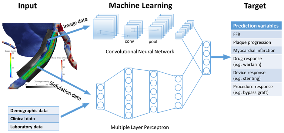
Machine LearningMachine learning has a growing role in medicine, but it has not reached to its potential, especially in cardiovascular imaging. We believe machine learning can be used to build predictive models and integrate data from all possible sources (imaging or non-imaging) by a hybrid approach based on conventional feature-based learning and the latest deep learning methods. We are working on the following projects:
|
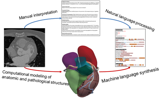
Natural language processingThe final task of performing a cardiovascular imaging procedure is to prepare a written report, which will be used to communicate the findings and clinical implications with the referring physician. Traditionally, these reports are written largely as free text and have inconsistence structure and terminology from site to site and from physician to physician. Therefore, structured reporting is increasingly recommended to assure quality and consistency. By analyzing the the existing reports using natural language processing, our goal is to search for knowledge and relationship between the reported and actual findings detected by image processing and to facilitate the structured reporting by automatic generation of key elements and paragraphs that can be faithfully extracted from the images.
|
|
|
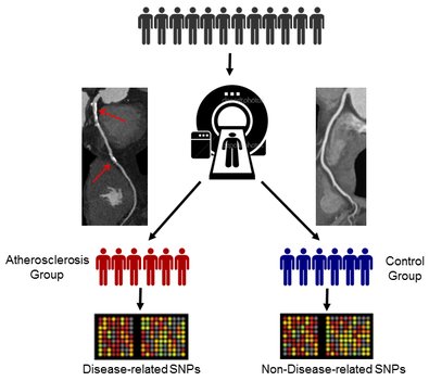
Imaging GenomicsCoronary artery disease is a common condition with a known heritable component that has spurred interest in genetic research for decades, resulting in a handful of candidate genes and an appreciation for the complexity of its genetic contributions. With the widespread use of high-throughput genotyping pipeline such as next-generation sequencing, combined with big data-driven solutions in bioinformatics, translating the emerging genetic technologies into clinical practice and, therefore, provide valuable insight into the study of coronary artery disease. Recently, noninvasive imaging technologies have dramatically improved our ability to detect subclinical atherosclerosis in a safe and reproducible manner in large numbers of patients. Our goal is to apply imaging to identify early-stage phenotypic markers in genetic association studies of coronary artery disease.
|
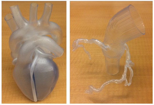
3D Printing3D printing has emerged as a promising method to address issues of traditional manufacturing strategies, with the ability to create virtually any geometry using a variety of biocompatible and soft materials that was heretofore infeasible or time-consuming. 3D printing is an additive manufacturing process that is performed by depositing layers of material sequentially according to a pre-designed digital model. New developments allow printing with multiple materials with different material properties like elasticity and porosity. With the increasing availability and reduced cost of 3D printing machines, this technology becomes a promising method to create physical replica of complex patient-specific cardiovascular structures constructed from 3D imaging modalities, e.g. CT, MR and echo. Our current research is to use 3D printing to fabricate patient-specific arterial models with realistic material properties and investigate flow and pressure in these models with respect to various upstream and downstream physiologic conditions. In addition, we are designing and fabricating new medical devices for structural heart diseases.
|
|
|
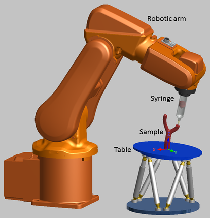
RoboticsThe overall scheme of 3D printing adopted in all open and commercial platforms is typically to first convert the desired object into layers of 2D slices and then deposit materials sequentially in a layer-by-layer fashion. When this method is used to form large structures of complex curved shapes, as they occur in natural cardiac tissues and blood vessels, three fundamental issues exist. One issue is the necessity of adding and removing support material to prevent overhanging features from collapsing into subsequent layers. Furthermore, thin-walled shell structures of high curvature are also difficult to fabricate since the printer head is only directed to move within a 2D plane and formation of the shell requires printing small pieces in many slices, which leads to serious artifacts of discretization. The last issue is the constraint of aligning internal structures to a Cartesian grid. Thus, it is impossible to create scaffolds mimicking curvilinear fiber orientations observed in all cardiovascular tissues. We are developing a new 3D printing system that utilizes a fluid dispensing nozzle mounted on a robotic arm and a rotary moving table. Our system is especially designed to fabricate curved scaffolds of any internal structure without supports.
|
